The new electron microscope enables work at cryo-conditions and provides higher resolution
At the beginning of September, the Laboratory of Electron Microscopy started operation of the new transmission electron microscope JEOL JEM-1400. The new microscope is not only capable of visualizing cellular ultrastructure, nanomaterials, isolated virions, bacteria, and protein macromolecules; but also offers a possibility to reconstruct the 3D volume of specimens (electron tomography).
The transmission electron microscope JEOL JEM 1400 replaced the 25 years old routine microscope JEOL JEM-1010. A great advantage against its predecessor is the possibility to work at cryo-conditions, allowed by the new cryo-tomography grid holder. Moreover, this microscope is equipped with a holder for four EM grids, which speeds up the work immensely. The movement of the holder is fully motorized, which allows to record the position of structure on the grid and recall it later. The users will also appreciate an autofocus button.
The new camera of the TEM provides better resolution (20 Mpix). Its software allows user-friendly, automated stitching of adjacent images (up to grid 5x5) to enlarge the view of an area of interest at the desired resolution.
Detailed specification of the new microscope JEOL JEM-1400 Flash:
Acceleration voltage: 80 – 120 kV
Source of electrons: heated tungsten wire / LaB6 cathode
Point resolution: 0.38 nm
Image acquisition: CMOS camera XAROSA (EMSIS BmbH) with resolution 20 Mpix
Other equipment: holder for up to 4 grids, grid holder for cryo-electron tomography, Hight-tilt grid holder for tomography (80 deg), software for image stitching and corelative microscopy
Employees and students can book the microscope through online booking system or by contact person.

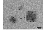
Carbon soot

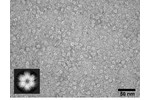
Particles of hexameric protein...

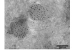
Immunolocalization of a protein...

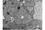
Ultrastructure of neurocytes.


